Looking back, Gary Morin, professor and chairman of the Department of Health and Movement Sciences, has mixed emotions about a cadaver-based anatomy course he completed as a college student. “I have to admit that I didn’t like it,” says Morin. “But I loved how much I learned.”
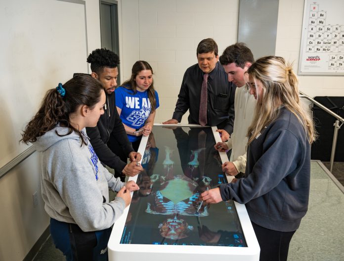
Today, he remains a firm believer in the power of experiential learning, forwarded by Southern’s recent acquisition of an Anatomage Table — a high-tech, 3D system for teaching anatomy and physiology as well as conducting virtual dissections. The table, which is housed in the new home of the College of Health and Human Services, features four life-size virtual cadavers: two females and two males.
The digital images are created from actual human cadavers, “meticulously segmented from photographic images to deliver the most accurate real 3D anatomy,” says medical technology supplier Anatomage. The result: extreme accuracy without chemicals, unpleasant smells, recurring costs, or regulations — and a much higher degree of student comfort compared with traditional cadavers.
“It allows us to teach so many things you can’t with a regular cadaver,” says Morin, of the table’s functionality. For example, students can view a digital “pregnancy,” including detailed fetal anatomy. They also can study movement by watching kinesiology simulations of a shoulder, knee, or hip — or examine the physiological functioning of a beating heart.
“It’s incredible to see what this technology can do and the excitement of the students who are using it,” says Morin, who used the table this summer in a graduate-level clinical anatomy course during which students conducted virtual dissections using styluses.
The technology is in place at some of the nation’s leading medical schools and institutions, including the Mayo Clinic in Jacksonville, Florida. At Southern, Morin foresees numerous applications. The Department of Communication Disorders might use it to review head and neck anatomy, for example.
The table is highly portable. Much like a hospital bed, it can be wheeled to different rooms. Additionally, the tabletop flips upward, placing the virtual cadaver in a standing position for easy viewing by an entire class. The virtual cadaver also can be projected onto a classroom screen. “Our students are going to get a great education working with it,” says Morin.
He notes the importance of hands-on learning while acknowledging the many challenges of studying anatomy with a regular cadaver, something that has never been done at Southern. “They are hard to move. They weigh a lot. If they are lying on their back, you can’t simply put them over on their belly to work,” says Morin, reflecting on his own college experience. “But with this device, you simply slide two fingers across the table and the body flips over — and the learning continues.”


